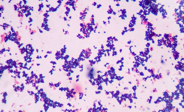
Have you ever wondered how scientists distinguish between the seemingly invisible world of bacteria? It’s a microscopic dance that involves a fascinating technique known as Gram staining, a cornerstone of microbiology. This technique, developed over a century ago, remains a critical tool for identifying and classifying bacteria. One of the most striking aspects of this technique is the distinct color change observed in bacteria, giving them a telltale identity. So, what color does gram-positive bacteria stain?

Image: pixelppt.blogspot.com
The answer is a vibrant **purple**. But why is this color so significant? It stems from the unique properties of the bacterial cell wall, a complex structure that defines the bacterium’s shape and plays a vital role in its survival. Let’s delve into the intricate world of Gram staining to understand why gram-positive bacteria take on this distinct purple hue.
The Magic of Gram Staining: A Journey into Bacterial Structures
Gram staining, named after its inventor, Hans Christian Gram, is a differential staining technique that exploits the structural differences between bacterial cell walls. It’s a simple yet powerful tool that allows microbiologists to categorize bacteria into two primary groups: gram-positive and gram-negative.
Understanding the Structure
The key lies in the intricate composition of the bacterial cell wall. The cell wall acts as a protective barrier, safeguarding the bacterium’s internal environment from external stresses. This sturdy structure is composed of peptidoglycan, a unique polymer made of sugars and amino acids. The amount and arrangement of peptidoglycan vary significantly between gram-positive and gram-negative bacteria.
Gram-Positive Bacteria: A Thick Peptidoglycan Layer
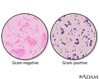
Gram-positive bacteria possess a thick layer of peptidoglycan that comprises nearly 90% of their cell wall. This thick layer acts like a robust fortress, providing structural support and resistance to various environmental challenges.

Image: ssl.adam.com
Gram-Negative Bacteria: A Thin Peptidoglycan Layer
In contrast, gram-negative bacteria have a much thinner layer of peptidoglycan, only about 10% of their cell wall. This thinner layer is sandwiched between an outer membrane and an inner membrane. This distinct structure gives gram-negative bacteria a somewhat more complex and resilient nature.
The Staining Process: A Step-by-Step Guide
Gram staining is a meticulous process that involves a series of steps. Each step plays a crucial role in revealing the secrets hidden within the bacterial cell structure.
1. Crystal Violet Staining: The First Step
The journey begins with the application of crystal violet, a primary stain that readily penetrates both gram-positive and gram-negative bacterial cell walls. The crystal violet molecules bind to the peptidoglycan, imparting a deep purple color to both types of bacteria.
2. Iodine Treatment: Forming a Complex
Next, iodine is added. This acts as a mordant, forming a complex with the crystal violet, creating a larger, more insoluble compound called a crystal violet-iodine complex. This complex becomes trapped within the thick peptidoglycan layer of gram-positive bacteria.
3. Decolorization: The Crucial Step
The crucial step in Gram staining is the decolorization step, usually with a solution of ethanol or acetone. The decolorizer disrupts the outer membrane of gram-negative bacteria, dissolving the thin peptidoglycan layer and releasing the crystal violet-iodine complex. However, the thick peptidoglycan layer of gram-positive bacteria prevents the decolorizer from penetrating effectively. The crystal violet-iodine complex remains trapped within their cell walls.
4. Safranin Counterstain: Distinguishing the Colors
The final step involves the application of a counterstain, typically safranin, a red dye. Safranin enters the cells that have been decolorized, staining them red. This step distinguishes the gram-negative bacteria, which now appear pink or red, from the gram-positive bacteria, which retain their original purple color.
The Significance of Gram Staining: A Window into the Microbial World
Gram staining, a seemingly simple technique, has revolutionized our understanding of bacteria. It’s a cornerstone of microbiology, providing insights into bacterial structure, allowing for rapid identification of bacterial species, and facilitating appropriate treatment strategies.
1. Rapid Identification: A Crucial Tool in Clinical Settings
The ability to quickly identify gram-positive and gram-negative bacteria is invaluable in clinical settings. This allows healthcare professionals to make informed decisions regarding patient care. By knowing the gram stain reaction, clinicians can promptly initiate targeted antibiotic therapy, significantly impacting patient outcomes.
2. Understanding Bacteria Structure: A Foundation for Research
Gram staining provides a vital entry point for studying bacterial structure. Understanding the unique features of gram-positive and gram-negative cell walls allows researchers to develop new antibiotics that specifically target these differences, leading to the development of more effective and targeted therapies.
3. Differentiating Between Bacteria: Guiding Treatment Strategies
Microbiologists use Gram staining to identify different bacterial species responsible for infections. This differentiation is crucial for selecting appropriate antibiotic therapy. Some antibiotics are effective against gram-positive bacteria, while others specifically target gram-negative bacteria. This tailored approach helps to ensure the most effective treatment and minimize the risk of antibiotic resistance.
4. Monitoring Infection: Assessing Treatment Success
Gram staining plays a vital role in monitoring the effectiveness of antibiotic treatment. The presence and distribution of gram-positive and gram-negative bacteria in clinical samples can provide valuable information about the progress of infection and the efficacy of ongoing therapy.
What Color Does Gram Positive Bacteria Stain
Gram Staining: Beyond Staining:
While Gram staining is a crucial technique for identifying bacteria, it’s important to remember that it’s not a standalone tool. Further investigations are often required to confirm an accurate identification. Additional tests, such as culturing, bio-molecular identification, and antibiotic susceptibility testing, provide a comprehensive picture of the bacterial infection.
Gram staining, a seemingly simple staining technique, holds a pivotal role in our understanding of the microbial world and its impact on human health. It’s a cornerstone of microbiology, providing a window into the intricate structures of bacteria, facilitating rapid identification, and guiding treatment strategies. The vibrant purple hue of gram-positive bacteria stands as a testament to the power and elegance of this technique.


/GettyImages-173599369-58ad68f83df78c345b829dfc.jpg?w=740&resize=740,414&ssl=1)


